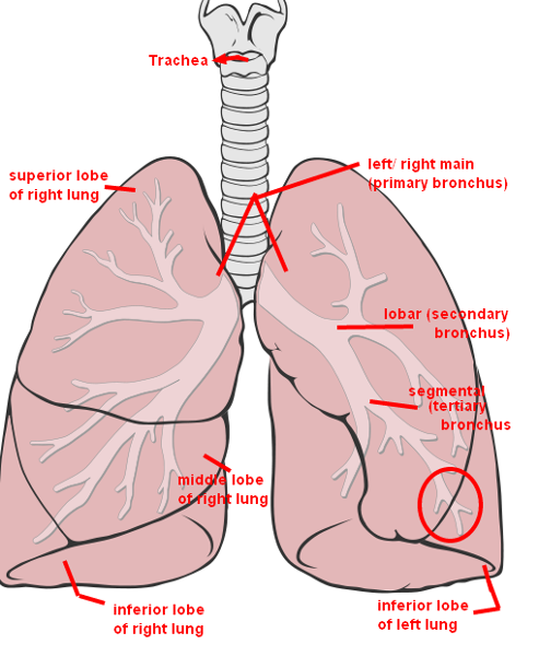
45 appropriately label all structures provided with leader lines on the model shown below
Neuromuscular junction: Parts, structure and steps - Kenhub The neuromuscular junction: Structure and function. At its simplest, the neuromuscular junction is a type of synapse where neuronal signals from the brain or spinal cord interact with skeletal muscle fibers, causing them to contract. The activation of many muscle fibers together causes muscles to contract, which in turn can produce movement ... Solved 8. Appropriately label all structures provided with | Chegg.com Appropriately label all structures provided with leader lines on the model shown below. 9. race a molecule of oxygen from the nostrils to the pulmonary capillaries of the lungs: Nostrils → ystem Structures 1. Complete the labeling of the model of the respiratory structures (sagittal section) shown below. 2.
A&P 139 Chapter 22 Reproductive System Flashcards - Quizlet true. The spermatogenic cells that line the seminiferous tubules produce sperm. Label the structures seen in this superior view of the female pelvis. (Note that the structures in the right half of the pelvis have been retracted). Complete the sentences describing the male external reproductive organs.

Appropriately label all structures provided with leader lines on the model shown below
Solved 8. Appropriately label all structures provided with | Chegg.com Question: 8. Appropriately label all structures provided with leader lines on the model shown below. This problem has been solved! See the answer Show transcribed image text Expert Answer Ans - 1- hyoid bone It is solitary u shaped bone situated in midline of neck. It is superior to thyroid cartilage. 2- thyrohyoid membran … View the full answer review sheet exercise 36 Flashcards - Easy Notecards review sheet exercise 36 Flashcards. label all structures provided with leader lines on the diagram below. label all structures provided with leader lines on the diagram below. label all structures provided with leader lines on the diagram below. Part 2. PDF Study Guide - Mr. Burghardt Ponderosa High School Shingle Springs, CA First, identify the structures in Column B by matchina them with the descrip- tions in Column A. Enter the correct letters (or terms if desired) in the answer blanks. Then, select a different color for each of the terms in Column B that has a color-coding circle and color in the structures on Fioure 6—2.
Appropriately label all structures provided with leader lines on the model shown below. PDF Lab 2: Endocrine Anatomy & Histology - IIS Windows Server the write-up. All images must be completely labeled with all of the structures listed in blue. Points will be lost for images that are not completely labeled. HINT: When we examined tissue types in API, students needed to use the highest power possible to focus on the small structures. In APII we will be looking at larger structures A & P II Lab Practical 2 Review Flashcards - Quizlet Appropriately label all structure provided with leader lines on the diagrams below. Lungs. ... For each of the following cases, check the column appropriate to your observations on the operation of the model lung. Change: In internal volume of the bell jar (thoractic cage) Review Sheet 36 - Anatomy of the Respiratory System - Quizlet Know and be able to label the following. ... The conducting zone structures; all respiratory passageways EXCLUDING the respiratory zone structures (alveolar sacs, alveolar ducts, respiratory bronchioles, alveoli, and respiratory membrane) ... Serous membranes a. line the mouth. b. have parietal and visceral layers. c. consist of epidermis and ... PDF The Cell: Anatomy and Division - Holly H. Nash-Rule, PhD In the following diagram, label all parts provided with a leader line. Nuclear envelope Nucleus Ribosomes Mitochondrion Peroxisome Golgi apparatus Rough endoplasmic reticulum Nuclear pore Nucleolus Cytosol Lysosome Centrioles Microtubule Intermediate filaments Microvilli Smooth endoplasmic reticulum Differences and Similarities in Cell Structure 5.
PDF Brain Anatomy - Wou BI 335 - Advanced Human Anatomy and Physiology Western Oregon University Figure 4: Mid-sagittal section of brain showing diencephalon (includes corpus callosum, fornix, and anterior commissure) Marieb & Hoehn (Human Anatomy and Physiology, 9th ed.) - Figure 12.10 Exercise 2: Utilize the model of the human brain to locate the following structures / landmarks for the PDF Document1 - Gore's Anatomy & Physiology 14. Relative to general terminology concerning muscle activity. first label the following structures on Figure 6—5: insertion. origin. tendon. resting mtyscle. and contracting muscle. Next, identify the two structures named below by choosing different colors for the coding circles and the corresponding structures in the figure. Movable bone BI217 Sec.750 Spring 2017: Exercise 36 Flashcards | Quizlet Appropriately label all structures provided with leader lines on the diagrams below. 9. Trace a molecule of oxygen from the nostrils to the pulmonary capillaries of the lungs: Nostrils → NOSTRILS -> NASAL CAVITY -> PHARYNX -> LARYNX -> TRACHEA -? Exercise 23 - Anatomy_Respiratory System.pdf - Course Hero Appropriately label all structures provided with leader lines on the model fluid? bronchi: passageways? shown below. Review Sheet 23 299 9. Match the terms in column B to those in column A. Column A 1. pleural layer attached directly to the lung 2. "floor" of the nasal cavity — 3. food and fluid passageway inferior to the laryngopharynx 4 ...
PDF Chapter 3 Review Materials Key - wtps.org Referring to plasma membranes, circle the term or phrase that does not aectrodes and leads Reading of -70 rnV on oscilloscope ining of dioestive trac belong in each of the following groupings. 1. Fused protein molecules of adjacent cells Communication between adjacent c Tight junction No intercellular space 2. Plasma Membrane Structure - Function, Components, Structure ... - BYJUS The model describes plasma membrane structure as a mosaic of components which includes proteins, cholesterol, phospholipids, and carbohydrates; it imparts a fluid character on the membrane. Thickness of the membrane is in the range of 5-10nm. The proportion of constituency of plasma membrane i.e., the carbohydrates, lipids and proteins vary ... PDF Practice Quiz Tissues •Lines ureters, bladder and part of the urethra. Identify the structure indicated. Loose Areolar Connective Tissue Elastic Fiber. ... •Provides strong attachment between structures that have Tissue forces pulling in one direction. Identify the tissue type and a location where it is found. Spongy Bone Tissue The Structure of DNA - University of Arizona The Structure of DNA The Structure of DNA This figure is a diagram of a short stretch of a DNA molecule which is unwound and flattened for clarity. The boxed area at the lower left encloses one nucleotide. Each nucleotide is itself make of three subunits: A five carbon sugar called deoxyribose (Labeled S)
Spinal Cord Cross Section Explained (with Videos ... - New Health Advisor The Structure. Exiting through a big hole at the bottom of the skull, the spinal cord is covered by the vertebral column that protects it. The spinal nerves come out from the spaces between the bony arches in pairs. They are named for the area of the vertebral column from which they come. These areas include: Cervical or neck; Thoracic or chest
Solved 8. Appropriately label all structures provided with - Chegg We review their content and use your feedback to keep the quality high. 100% (10 ratings) The larynx is a tough and flexible segment of the respiratory tract connecting the pharynx to the trachea of …. View the full answer. Transcribed image text: 8. Appropriately label all structures provided with leader lines on the model shown below.
4.1 Types of Tissues - Anatomy & Physiology Tissue Membranes. A tissue membrane is a thin layer or sheet of cells that either covers the outside of the body (e.g., skin), lines an internal body cavity (e.g., peritoneal cavity), lines a vessel (e.g., blood vessel), or lines a movable joint cavity (e.g., synovial joint). Two basic types of tissue membranes are recognized based on the primary tissue type composing each: connective tissue ...


Post a Comment for "45 appropriately label all structures provided with leader lines on the model shown below"