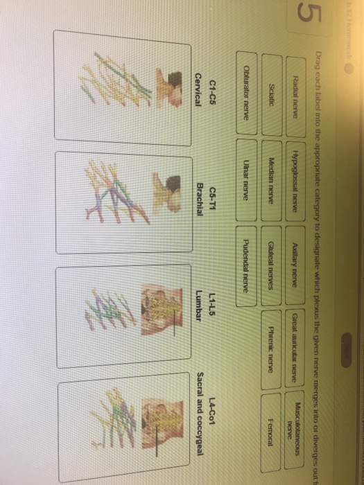
41 drag each label into the appropriate category to identify from which plexus the given nerve emerges
Chapter 13 Worksheet Flashcards - Quizlet Drag each label to accurately identify the regions of spinal nerves. Drag each label into the appropriate category to designate whether the given item describes elements of gray or white matter of the spinal cord. Sets found in the same folder CBIO Figures 20 terms KLC38718 PLUS Chapter 3 Worksheet 50 terms sonjamilosavljevic Chapter 14 Worksheet 12 pairs of cranial nerves: What are they and what are their ... - CogniFit Classification of the 12 pairs of cranial nerves Each cranial nerve is paired and is present on both sides. There are twelve cranial nerves pairs, which are assigned Roman numerals I-XII, sometimes also including cranial nerve zero. There are XII cranial nerves on the left hemisphere of the brain and exactly the same on the right hemisphere.
"Anatomy and Physiology Lab I" on OpenALG Label each of the following drawings of cells in different stages of mitosis and cytokinesis. Lesson 3: Histology - Epithelial & Connective Tissues . Created by Dan McNabney. Introduction . In this lesson you will describe what a tissue is, define each of the four primary tissues types, and explore epithelial and connective tissues in detail.
Drag each label into the appropriate category to identify from which plexus the given nerve emerges
Cervical plexus: Anatomy, branches, course, innervation - Kenhub The cervical plexus is formed in the neck region. It lies deep to the sternocleidomastoid muscle, and anterolateral to the levator scapulae. Each of the cervical nerves forming the plexus communicates with one another in a superior-inferior fashion close to their origins, thus C2 accepts communicating fibres from C1, C3 from C2, and so on. Nervous System Chapter 13 Flashcards - Quizlet -Somas, synapses, and dendrites Gray matter Drag each label into the appropriate category to designate whether the given item describes elements of gray or white matter of the spinal cord. -Contains myelinated axons -Located to the periphery of the spinal cord -Transmits electrical signals rapidly over long distances White matter AHCDW9Notes5.pdf - 5. Award: 10.00 points Problems? Adjust... Drag each label into the appropriate category to identify from which plexus the given nerve emerges. Explanation: Plexus is a branching and diverging network of nerve roots. The five plexuses are the cervical, brachial, lumbar, sacral, and coccygeal.
Drag each label into the appropriate category to identify from which plexus the given nerve emerges. Drag each label into the appropriate category to | Chegg.com Drag each label into the appropriate category to identify from which plexus the given nerve emerges; Question: Drag each label into the appropriate category to identify from which plexus the given nerve emerges. This question hasn't been solved yet What are the 12 cranial nerves? Functions and diagram Each nerve has a name that reflects its function and a number according to its location in the brain. Scientists use Roman numerals from I to XII to label the cranial nerves in the brain. The 12... Full text of "Human Anatomy, 6 edition" - Internet Archive An icon used to represent a menu that can be toggled by interacting with this icon. The appropriate use of neurostimulation of the spinal cord and ... The Neuromodulation Appropriateness Consensus Committee (NACC) of the International Neuromodulation Society (INS) evaluated evidence regarding the safety and efficacy of neurostimulation to treat chronic pain, chronic critical limb ischemia, and
(DOC) HUMAN BRAIN AND PLANT TISSUE ELECTRIFICATION ... - Academia.edu Electrical stimulation of various sites of the Cell Membrane of Ariny Amos triggers electric signals transmitted to the central Nervous system of the Human Brain systems in action potential as electric signals effects on the Hypothalamus, The Spinal Cord | Boundless Anatomy and Physiology - Course Hero The spinal cord is a long, thin, tubular bundle of nervous tissue and support cells that extends from the medulla oblongata of the brain to the level of the lumbar region. The brain and spinal cord together make up the central nervous system (CNS). The spinal cord, protected by the vertebral column, begins at the occipital bone and extends down ... Biological Psychology , Ninth Edition - SILO.PUB Each chapter is divided into modules; each module begins with its own introduction and finishes with its own summary and questions. This organization makes it easy for instructors to assign part of a chapter per day instead of a whole chapter per week. Solved drag each label into the appropriate category to | Chegg.com drag each label into the appropriate category to designate which plexus the given nerve merged into or diverges out from Show transcribed image text Expert Answer 100% (13 ratings) Answere: Please drag the corresponding labes of nerves to correspomding plexus as suggested below. C1-C5 Cer … View the full answer
Lymphatic system - Wikipedia The lymphatic system, or lymphoid system, is an organ system in vertebrates that is part of the immune system, and complementary to the circulatory system.It consists of a large network of lymphatic vessels, lymph nodes, lymphatic or lymphoid organs, and lymphoid tissues. The vessels carry a clear fluid called lymph (the Latin word lympha refers to the deity of fresh water, "Lympha") back ... Solved Drag each label into the appropriate category to | Chegg.com Drag each label into the appropriate category to identify from which plexus the given nerve emerges. Great auricular nerve Musculocutaneous nerve Sciatic nerve Obturator nerve Phrenic nerve Femoral nerve Axillary nerve Radial nerve Pudendal nerve Ulnar nerve Cervical Brachial Lumbar Sacral and Coccygeal 09 Human Anatomy & Physiology Laboratory Manual Main Version 10th Edition ... Correctly identify each of the nine regions of the abdominopelvic cavity by inserting the appropriate term for each of the letters indicated in the drawing. a. _____ b. ... Cells fall into four different categories according to their structures and functions. Each of these corresponds to one of the four tissue types: epithelial, muscular ... chapter 14 a&p Flashcards | Quizlet Drag each label into the appropriate category to designate which plexus the given nerve merges into or diverges out from. c1 -c5 cervical -hypoglossal nerve -phrenic nerve c5-t1 brachial -axillary nerve -musculocutaneous nerve l1-l5 lumbar -femoral -obturator nerve l4-Co1 sacral and coccygeal -sciatic -gluteal nerves
Full text of "NEW" - Internet Archive An icon used to represent a menu that can be toggled by interacting with this icon.
Spinal Nerves | Boundless Anatomy and Physiology - Course Hero Each spinal nerve is formed by the combination of nerve fibers from the dorsal and ventral roots of the spinal cord. The dorsal roots carry afferent sensory axons, while the ventral roots carry efferent motor axons. The spinal nerve emerges from the spinal column through an opening (intervertebral foramen) between adjacent vertebrae.
Divisions of the Autonomic Nervous System - Course Hero The autonomic nervous system regulates many of the internal organs through a balance of two aspects, or divisions. In ition to the endocrine system, the autonomic nervous system is instrumental in homeostatic mechanisms in the body. The two divisions of the autonomic nervous system are the sympathetic division and the parasympathetic division.
Cranial Nerves List And Their Functions - Byju's Cranial nerves are basically named according to their structure and functions. Olfactory and optic nerves emerge from the cerebrum and all other 10 nerves emerge from the brain stem. Cranial nerve functions are involved with the functioning of all five senses organs and muscle movements.
Chapter 13 and 14 Flashcards - Quizlet Identify the cerebral lobes on the left side of the figure. Label the additional cerebral structures on the right side of the figure. ... Drag each label into the appropriate category to designate which plexus the given nerve merges into or diverges out from. Match the type of reflex with its description. 1. The simplest reflex; muscles ...
Drag each label into the appropriate category to identify fr Academic Writing Biology Drag each label into the appropriate category to identify fr Format and Features Approximately 275 words/page All paper formats (APA, MLA, Harvard, Chicago/Turabian) Font 12 pt Arial/ Times New Roman Double and single spacing Free bibliography page Free title page 1 inch margin on all sides
CBIO Exam 5 Flashcards | Quizlet Drag each label into the appropriate category to identify from which plexus the given nerve emerges. Correctly identify and label the structures associated with the anatomy of a ganglion. Correctly identify and label the structures associated with the branches of the spinal nerve in relation to the spinal cord.
AHCDW9Notes4.pdf - 4. Award: 10.00 points Problems ... - Course Hero Adjust credit for all students. Drag each label into the appropriate category to designate whether the given item describes ascending or descending neural tracts. Explanation: The white matter regions of the spinal cord form three large regions known as the posterior, lateral, and anterior column.
AHCDW9Notes5.pdf - 5. Award: 10.00 points Problems? Adjust... Drag each label into the appropriate category to identify from which plexus the given nerve emerges. Explanation: Plexus is a branching and diverging network of nerve roots. The five plexuses are the cervical, brachial, lumbar, sacral, and coccygeal.
Nervous System Chapter 13 Flashcards - Quizlet -Somas, synapses, and dendrites Gray matter Drag each label into the appropriate category to designate whether the given item describes elements of gray or white matter of the spinal cord. -Contains myelinated axons -Located to the periphery of the spinal cord -Transmits electrical signals rapidly over long distances White matter
Cervical plexus: Anatomy, branches, course, innervation - Kenhub The cervical plexus is formed in the neck region. It lies deep to the sternocleidomastoid muscle, and anterolateral to the levator scapulae. Each of the cervical nerves forming the plexus communicates with one another in a superior-inferior fashion close to their origins, thus C2 accepts communicating fibres from C1, C3 from C2, and so on.


:watermark(/images/watermark_only_sm.png,0,0,0):watermark(/images/logo_url_sm.png,-10,-10,0):format(jpeg)/images/anatomy_term/gracile-fasciculus-1/IgGqZ5xfdHvCthGXZdl4cw_gracile_fasciculus.png)

Post a Comment for "41 drag each label into the appropriate category to identify from which plexus the given nerve emerges"