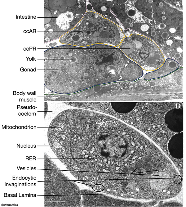
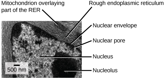
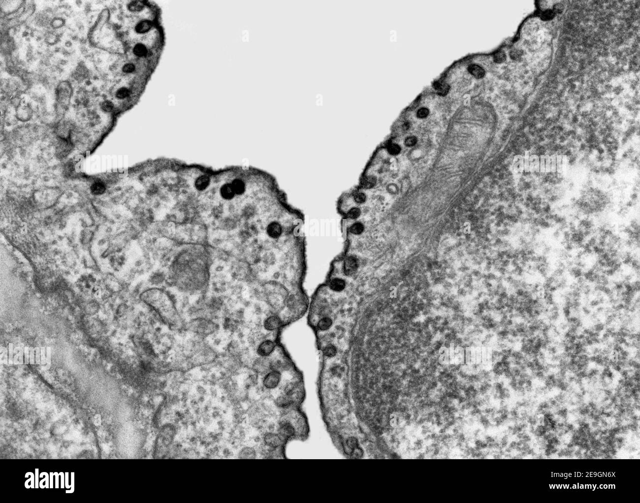
45 label the transmission electron micrograph of the cell
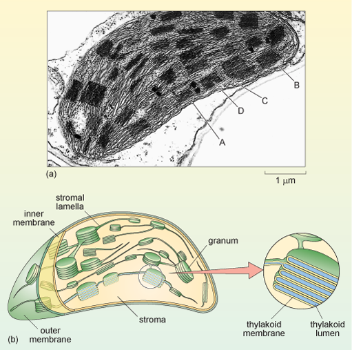
Solved Label the transmission electron micrograph of the | Chegg.com Transcribed image text: Label the transmission electron micrograph of the cell. 0 Nucleus rences Mitochondrion Heterochromatin Peroxisome Vesicle ULAR bumit Click and drag each label into the correct category to indicate whether it pertains to the cytoplasm or the plasma membrane. ICF Contacts the ECF Made of proteins and lipids Surrounds the cell Contains lon channels Organelles Fibers ... Label This Transmission Electron Micrograph : TEM of chloroplast from ... Label the transmission electron micrograph of the nucleus. Label the transmission electron micrograph of the nucleus. Transmission electron microscopy (tem) is a microscopy technique in which a beam of electrons is transmitted through a specimen to form an image. Figures label this transmission electron micrograph ( 16, . CIN2003. Ian Roberts.
A&P Unit 2 Exam Flashcards | Quizlet Cells in a lymph node that engulf and destroy damaged cells, foreign substances, and cellular debris are ... Label the transmission electron micrograph based on the hints provided. Plasma cells. produce antibody molecules. Name the cells included in the mononuclear phagocytic system. macrophages monocytes

Label the transmission electron micrograph of the cell
Lap Practical #1 EC Flashcards | Quizlet Label the transmission electron micrograph of the cell. Fill-in the appropriate description with the correct type of cartilage. Elastic cartilage is composed of a network of branching elastic fibers. Figure 2. [Transmission electron micrograph of the...]. - Webvision ... Transmission electron micrograph of the RPE cells and the RPE-choroid interface in a normal human donor eye. cc, choriocapillaris; BM, Bruchs membrane; RPE, retinal pigment epithelium; ROS, rod photoreceptor outer segments; Ph, phagosomes; PG, pigment granules; TJ, tight junctions. Native Immunogold Labeling of Cell Surface Proteins and Viral ... Cells on Aclar disks were washed twice with PBS and then fixed with 2.5% glutaraldehyde in 0.1 M phosphate buffer (pH 7.4) for conventional transmission electron microscopy examination. Cells on the gold Quantifoil TEM grids were plunge-frozen immediately after the final wash with culture medium.
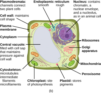
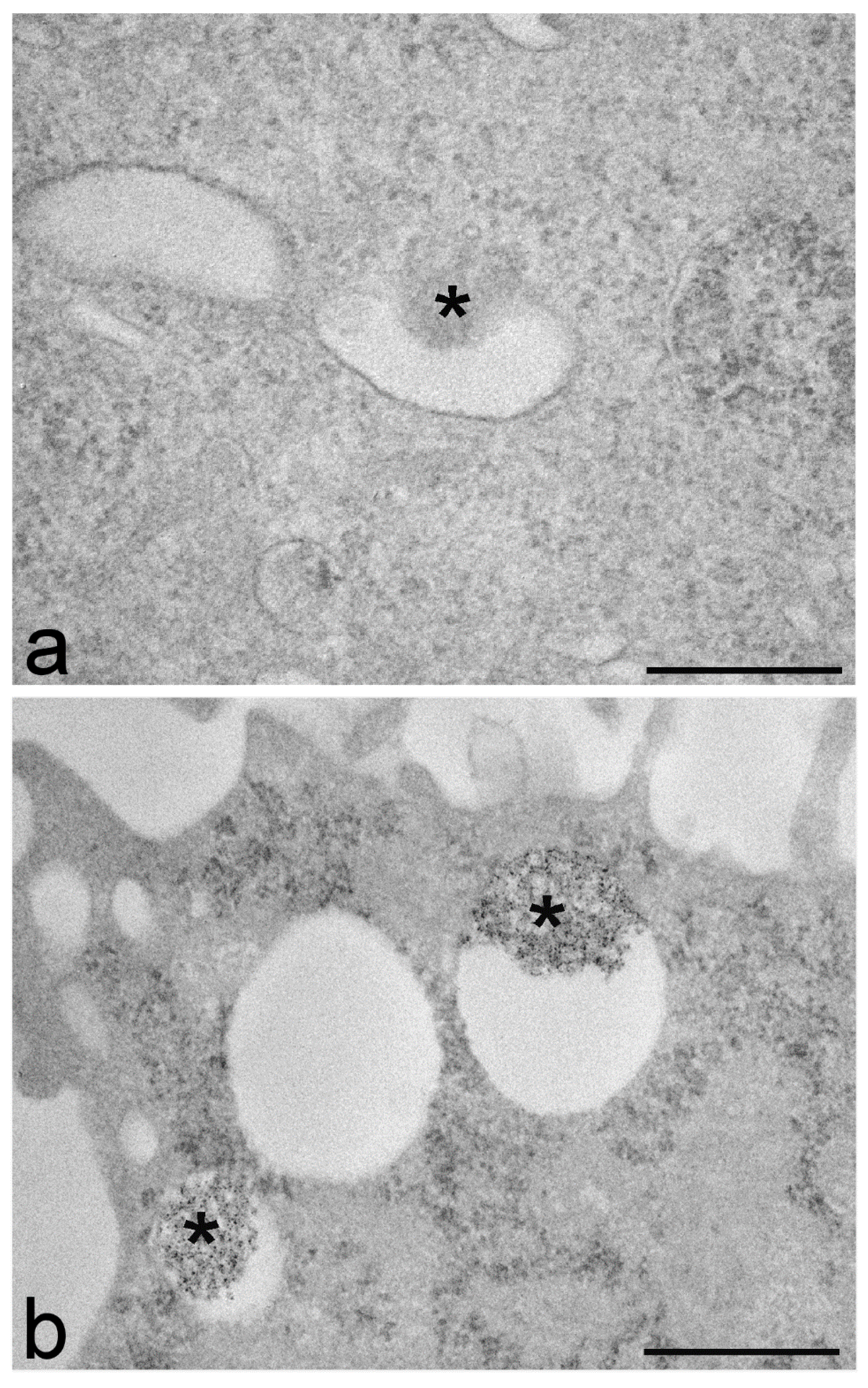
Label the transmission electron micrograph of the cell. PDF Identifying Organelles from an Electron Micrograph The electron micrograph displayed below illustrates many of the plant cell characteristics discussed The cell wall, large central vacuole and chloroplasts are clearly visible Also visible is the clearly defined nucleus containing chromatin Nucleus Chromatin The vacuole in this mature plant cell from a leaf is large, and occupies about 80% of Transmission electron micrograph of cultured mouse mammary cell labeled ... Download scientific diagram | Transmission electron micrograph of cultured mouse mammary cell labeled as shown in Fig. 3; a thick section was used to increase the length of TMV label included in ... Visualizing endocytotic pathways at transmission electron microscopy ... PKH26 is a fluorescent dye specific for long-lasting cell membrane labelling which has been successfully used for investigating cell internalization processes, at either flow cytometry or fluorescence microscopy. In the present work, diaminobenzidine photo-oxidation was tested as a procedure to detect PKH26 dye at transmission electron microscopy. Transmission Electron Micrograph of transfected HL-1 cells labeled for ... A and B. Single immunogold labeling experiments used 15 nm gold particles to label GFP. A. Immunogold-labeled TMEM43-WT cells. The label (large dot) is associated with the endoplasmic reticulum ...
The Transmission Electron Microscope | CCBER Transmission electron microscopes (TEM) are microscopes that use a particle beam of electrons to visualize specimens and generate a highly-magnified image. TEMs can magnify objects up to 2 million times. In order to get a better idea of just how small that is, think of how small a cell is. It is no wonder TEMs have become so valuable within the ... Transmission Electron Microscope (With Diagram) Transmission Electron Microscope (With Diagram) In this article we will discuss about the design of transmission electron microscope, explained with the help of a diagram. In TEM a finely focused beam of electrons from an electron gun is passed through a specially prepared ultra thin section of the specimen. The beam is focused on a small area ... Solved Label the transmission electron micrograph based on - Chegg Transcribed image text: Label the transmission electron micrograph based on the hints provided Mitochondrion Heterochromatin Plasma cell Nucleus Rough endoplasmic reticulum Nucleolus Previous question Next question Label the transmission electron micrograph of the cell. 0 Nucleus ... Label the transmission electron micrograph of the cell. 0 Nucleus rences Mitochondrion Heterochromatin Peroxisome Vesicle ULAR bumit Click and drag each label into the correct category to indicate whether it pertains to the cytoplasm or the plasma membrane. ICF Contacts the ECF Made of proteins and lipids Surrounds the cell Contains lon ...
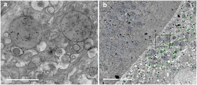
Transmission electron micrograph of U20S cells (whole mount) after ... Download scientific diagram | Transmission electron micrograph of U20S cells (whole mount) after labeling with mAb J143 and goat anti-mouse IgG coupled to 40/zm gold. The rectangular frame (frame ... Solved Label the transmission electron micrograph of the | Chegg.com 100% (4 ratings) Explanation - Mitochondrion is filamentous or globular in shape, occur in variable numbers from a few hundred to few thousands in different cells. It …. View the full answer. Transcribed image text: Label the transmission electron micrograph of the mitochondrion. Matrix granule Mitochondrion Outer membrane Cristae Inner ... BIO 224: Lab Midterm Review Flashcards - Quizlet Label the transmission electron micrograph of the mitochondrion. Synthesizes protein for secretion, insertion into the plasma membrane, and lysosomal enzymes. ... Label the transmission electron micrograph of the cell. proton. What is the name of the positively charged subatomic particle? Transmission electron micrograph of a cell loaded with magnetic ... Download scientific diagram | Transmission electron micrograph of a cell loaded with magnetic nanoparticles, which are confined in endosomes or lysosomes. from publication: Cell labeling with ...
CiteSeerX — Citation Query Quantization of L-Selectin distribution on ... Quantization of L-Selectin distribution on human leukocyte microvilli by immunogold labeling and electron microscopy," (1996) by R E Bruehl, T A Springer, D F Bainton Venue: J Histochem Cytochem., Add To MetaCart ... the leukocyte is a viscoelastic cell with the nucleus located in the intracellular space and cylindrical microvilli distributed ...
Electron Micrographs of Cell Organelles | Zoology It is an electron micrograph of cell's largest and most important organelle - the mitochondria and is characterized by the following features (Fig. 7 & 8): (1) The name mitochondria was given by Benda (1898) and their ma n function was brought to light by Kingsbury (1912). (2) Each mitochondria in section appears as sausage or cup or bowl ...
Study Chapter 14 & 15 Flashcards Flashcards - Quizlet Viruses and self-proteins are examples of proteins produced inside of the cell. True. ... Label the transmission electron micrograph based on the hints provided. Label the transmission electron micrograph based on the hints provided. Place the following tonsils in order based on their location from superior to inferior.
anatomy 10.png - Label the transmission electron micrograph of the ... anatomy 10.png - Label the transmission electron micrograph of the. anatomy 10.png - Label the transmission electron micrograph... School Utah Valley University; Course Title ZOOL 1090; Uploaded By emileeroylance19. Pages 1 Ratings 67% (3) 2 out of 3 people found this document helpful;
Label the transmission electron micrograph of the nucleus. Label the transmission electron micrograph of the cell. 0 Nucleus rences Mitochondrion Heterochromatin Peroxisome Vesicle ULAR bumit Click and drag each label into the correct category to indicate whether it pertains to the cytoplasm or the plasma...
Transmission electron micrograph of epidermal Langerhans cells ... Download scientific diagram | Transmission electron micrograph of epidermal Langerhans cells. (x89,000.) Colocalization of BL6-AU 15 and of BL2-AU 5 (arrows) in the same Birbeck granules. (A) The ...
Transmission Electron Microscope (TEM)- Definition, Principle, Images The working principle of the Transmission Electron Microscope (TEM) is similar to the light microscope. The major difference is that light microscopes use light rays to focus and produce an image while the TEM uses a beam of electrons to focus on the specimen, to produce an image. Electrons have a shorter wavelength in comparison to light which ...
Labeling the Cell Flashcards | Quizlet Label the transmission electron micrograph of the cell. ... Label the transmission electron micrograph of the nucleus. membrane bound organelles. golgi apparatus, mitochondrion, lysosome, peroxisome, rough endoplasmic reticulum. nonmembrane bound organelles. ribosomes, centrosome, proteasomes.
Transmission electron microscopy DNA sequencing - Wikipedia Transmission electron microscopy DNA sequencing is a single-molecule sequencing technology that uses transmission electron microscopy techniques. The method was conceived and developed in the 1960s and 70s, but lost favor when the extent of damage to the sample was recognized. In order for DNA to be clearly visualized under an electron microscope, it must be labeled with heavy atoms.
Label This Transmission Electron Micrograph / Microscopy ... - Blogger The applicability and strength of mettem is demonstrated by a study. Interpretation of electron micrographs to identify organelles and deduce the functions of specialized cells. Label the transmission electron micrograph of the nucleus. Label the transmission electron micrograph of the cell. Label the transmission electron micrograph of the.
Native Immunogold Labeling of Cell Surface Proteins and Viral ... Cells on Aclar disks were washed twice with PBS and then fixed with 2.5% glutaraldehyde in 0.1 M phosphate buffer (pH 7.4) for conventional transmission electron microscopy examination. Cells on the gold Quantifoil TEM grids were plunge-frozen immediately after the final wash with culture medium.
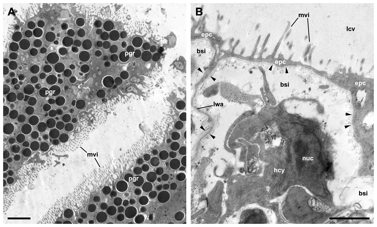
Figure 2. [Transmission electron micrograph of the...]. - Webvision ... Transmission electron micrograph of the RPE cells and the RPE-choroid interface in a normal human donor eye. cc, choriocapillaris; BM, Bruchs membrane; RPE, retinal pigment epithelium; ROS, rod photoreceptor outer segments; Ph, phagosomes; PG, pigment granules; TJ, tight junctions.
Lap Practical #1 EC Flashcards | Quizlet Label the transmission electron micrograph of the cell. Fill-in the appropriate description with the correct type of cartilage. Elastic cartilage is composed of a network of branching elastic fibers.




















Post a Comment for "45 label the transmission electron micrograph of the cell"