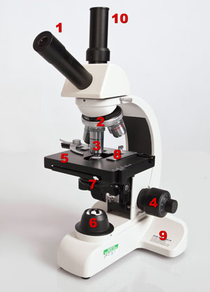
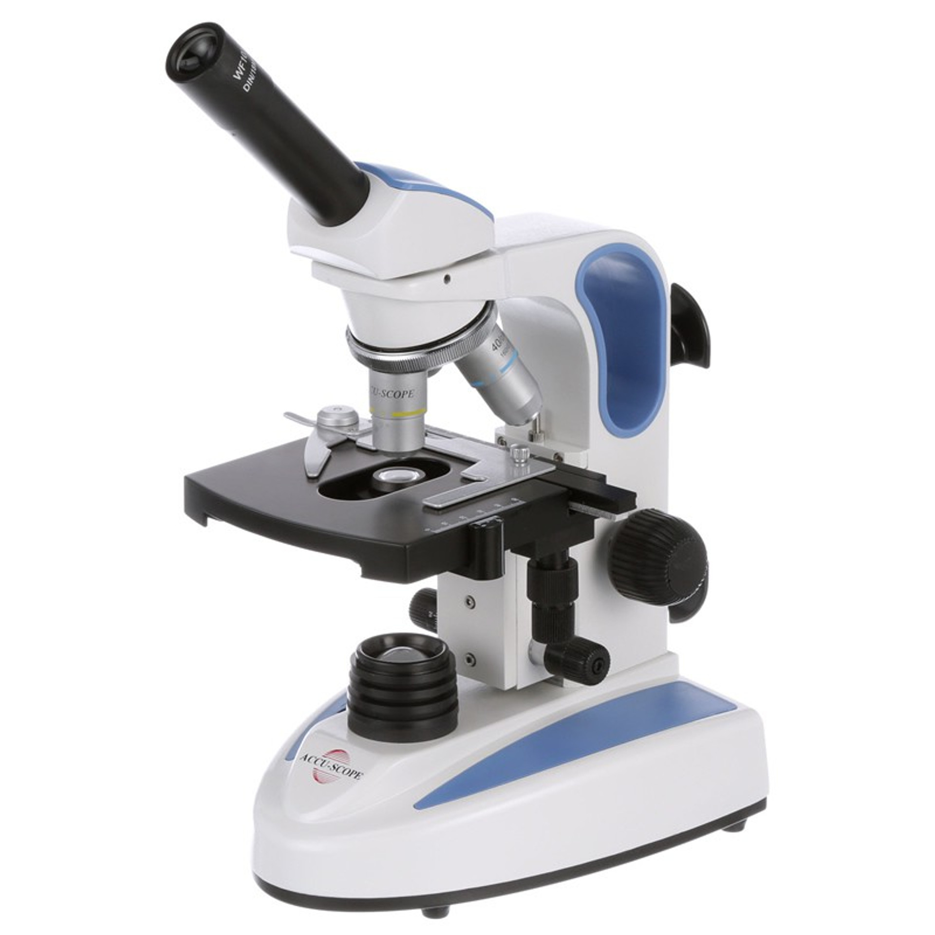
41 microscope labeled diagram
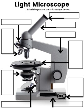
Parts of a microscope with functions and labeled diagram 17/09/2022 · 82 thoughts on “Parts of a microscope with functions and labeled diagram” Rolenzo. September 30, 2022 at 3:32 AM . This really good it helps us in our academicks. Reply. watty. September 27, 2022 at 11:04 PM . it is really helpful giving even some self testing questions. Reply. Shadow fighter. September 18, 2022 at 7:39 PM . this website has been very … Label the microscope — Science Learning Hub All microscopes share features in common. In this interactive, you can label the different parts of a microscope. Use this with the Microscope parts activity to help students identify and label the main parts of a microscope and then describe their functions. Drag and drop the text labels onto the microscope diagram.
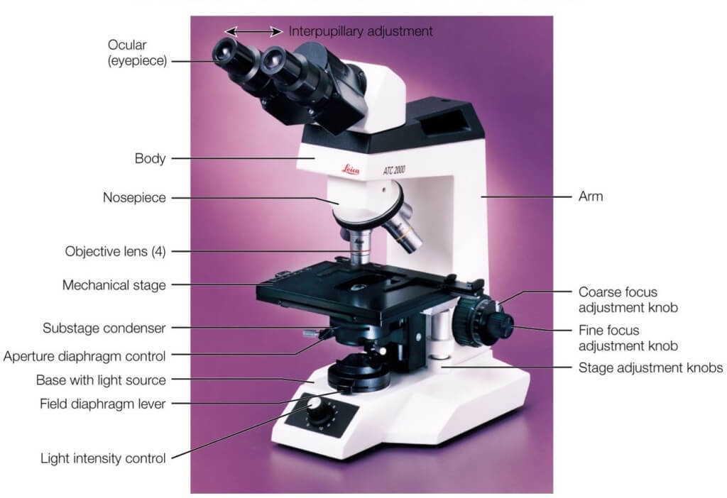
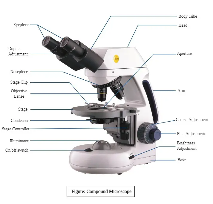
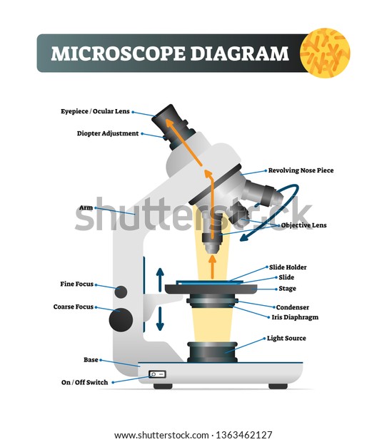
Compound Microscope Parts - Labeled Diagram and their Functions Labeled diagram of a compound microscope Major structural parts of a compound microscope There are three major structural parts of a compound microscope. The head includes the upper part of the microscope, which houses the most critical optical components, and the eyepiece tube of the microscope.
Microscope labeled diagram
Duck Anatomy - External and Internal Features with Labeled ... Jul 24, 2021 · Here, I will show you again the duck anatomy diagram as a whole so that you may summarize your contents so quickly. I tried to show you the basic anatomical features of a duck. If you need more duck labeled diagrams, please let me know. Or, you may join to anatomy learner on social media to get more updates on duck labeled diagrams. Fluorescence Microscopy- Definition, Principle, Parts, Uses 12/04/2022 · Light Microscope- Definition, Principle, Types, Parts, Labeled Diagram, Magnification; Parts of a microscope with functions and labeled diagram; Organic waste recycling (methods, steps, significance, barriers) Limitations of Fluorescence Microscope. Fluorophores lose their ability to fluoresce as they are illuminated in a process called … Cell Nucleus – function, structure, and under a microscope [In this figure] The cell nucleus diagram and its structure. The nucleus consists of the nuclear envelope like double-layer membranes with pores ( nuclear pores), DNA, nucleolus (a site for ribosome synthesis, plural: nucleoli ), nucleoplasm (like cytoplasm of a cell), and the nuclear matrix, a supportive structure like the cytoskeleton supports cells.
Microscope labeled diagram. Cat Skeleton Anatomy with Labeled Diagram - AnatomyLearner May 29, 2021 · Cat skeleton anatomy labeled diagram. Now, I will show you all the bones from the cat skeleton with a diagram. If you find any mistakes in this cat anatomy labeled diagram, please let me know. I hope this cat skeletal system anatomy labeled diagram might help you understand and identify all the cat’s bones. Parts of Stereo Microscope (Dissecting microscope) – labeled ... Labeled part diagram of a stereo microscope Major structural parts of a stereo microscope. There are three major structural parts of a stereo microscope. The viewing Head includes the upper part of the microscope, which houses the most critical optical components, including the eyepiece, objective lens, and light source of the microscope. Simple Microscope - Diagram (Parts labelled), Principle, Formula and Uses Parts of a Simple Microscope A simple microscope consists of Optical parts Mechanical parts Labeled Diagram of simple microscope parts Optical parts The optical parts of a simple microscope include Lens Mirror Eyepiece Lens A simple microscope uses biconvex lens to magnify the image of a specimen under focus. compact bone diagram microscope Histology bone compact tissue connective slides human lamellae bones diagram structure anatomy cartilage ground section biology tooth bio physiology ou. Bone section structure compact histology cross cortical function tissue animal ligaments edu tendons wsu mechanical practical anatomy. Anatomy & physiology i bis 240: osteon-canaliculi
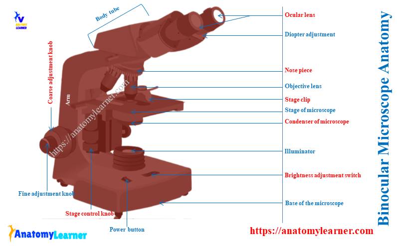
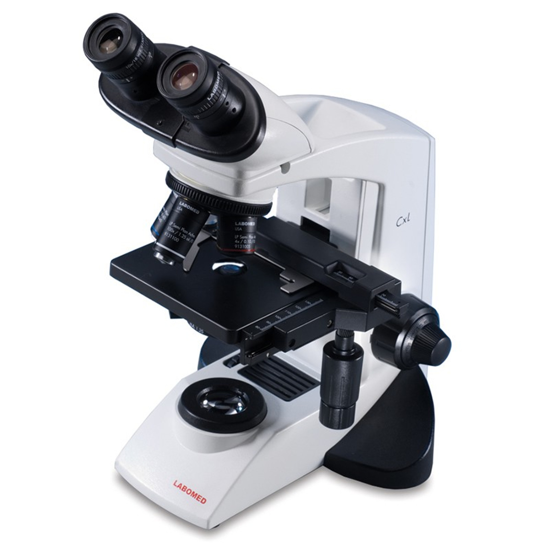
Microscope Parts, Function, & Labeled Diagram - slidingmotion Microscope parts labeled diagram gives us all the information about its parts and their position in the microscope. Microscope Parts Labeled Diagram The principle of the Microscope gives you an exact reason to use it. It works on the 3 principles. Magnification Resolving Power Numerical Aperture. Parts of Microscope Head Base Arm Eyepiece Lens Microscope Types (with labeled diagrams) and Functions Simple microscope labeled diagram Simple microscope functions It is used in industrial applications like: Watchmakers to assemble watches Cloth industry to count the number of threads or fibers in a cloth Jewelers to examine the finer parts of jewelry Miniature artists to examine and build their work Also used to inspect finer details on products Parts of a microscope with functions and labeled diagram Figure: Diagram of parts of a microscope There are three structural parts of the microscope i.e. head, base, and arm. Head - This is also known as the body. It carries the optical parts in the upper part of the microscope. Base - It acts as microscopes support. It also carries microscopic illuminators. Binocular Microscope Anatomy - Parts and Functions with a Labeled Diagram Now, I will discuss the details anatomy of the light compound microscope with the labeled diagram. Why it is called binocular: because it has two ocular lenses or an eyepiece on the head that attaches to the objective lens, this ocular lens magnifies the image produced by the objective lens. Binocular microscope parts and functions
Sperm Under Microscope with Labeled Diagram - AnatomyLearner Sperm Under Microscope 400X Labeled Diagram Before that, you may also read the below-mentioned article to get a full idea of the structure of seminiferous tubules - Histological features of the seminiferous tubules with the labeled diagram Okay, first, let's see the different histological features of the seminiferous tubules of an animal. Microscope: Parts Of A Microscope With Functions And Labeled Diagram. Figure: A diagram of a microscope's components. The microscope has three basic components: the head, the base, and the arm. Head:Occasionally, the head is considered the body. It holds the optical components of the upper part of the microscope. Base:The microscope's base provides great support. Interactive Bacteria Cell Model - CELLS alive Ribosomes: Ribosomes give the cytoplasm of bacteria a granular appearance in electron micrographs.Though smaller than the ribosomes in eukaryotic cells, these inclusions have a similar function in translating the genetic message in messenger RNA into the production of peptide sequences (proteins). Compound Microscope- Definition, Labeled Diagram, Principle, … 03/04/2022 · Magnification of compound microscope. In order to ascertain the total magnification when viewing an image with a compound light microscope, take the power of the objective lens which is at 4x, 10x or 40x and multiply it by the power of the eyepiece which is typically 10x.
Microscope labeled diagram - SlideShare Microscope labeled diagram 1. The Microscope Image courtesy of: Microscopehelp.com Basic rules to using the microscope 1. You should always carry a microscope with two hands, one on the arm and the other under the base. 2. You should always start on the lowest power objective lens and should always leave the microscope on the low power lens ...
Duck Anatomy - External and Internal Features with Labeled Diagram ... 24/07/2021 · Excellent, here I will show you both the external and internal organs anatomy of a duck with a labeled diagram. You will get a list of interesting anatomical facts of a duck so that you may compare it with other poultry species. First, I will introduce the different body parts of a duck with you. Later, I will describe the essential internal ...
Labelled Diagram of Compound Microscope The below mentioned article provides a labelled diagram of compound microscope. Part # 1. The Stand: The stand is made up of a heavy foot which carries a curved inclinable limb or arm bearing the body tube. The foot is generally horse shoe-shaped structure (Fig. 2) which rests on table top or any other surface on which the microscope in kept.
PDF Label parts of the Microscope Label parts of the Microscope: . Created Date: 20150715115425Z
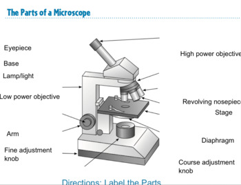
Parts of the Microscope Label and Definition Diagram | Quizlet Medium Power Objective. Provides magnification, usually about 10x; total magnification is 100. High Power Objective. Provides magnification, usually about 40x; total magnification is 400. Stage Clips. Grip slide in place for. viewing. Diaphragm. Controls amount of light entering the body tube.
Compound Microscope- Definition, Labeled Diagram, Principle ... The naked eye can now view the specimen at magnification 400 times greater and so microscopic details are revealed. Alternatively, the magnification of the compound microscope is given by: m = D/ fo * L/fe where, D = Least distance of distinct vision (25 cm) L = Length of the microscope tube fo = Focal length of the objective lens
A Study of the Microscope and its Functions With a Labeled Diagram May 21, 2019 - To better understand the structure and function of a microscope, we need to take a look at the labeled microscope diagrams of the compound ...
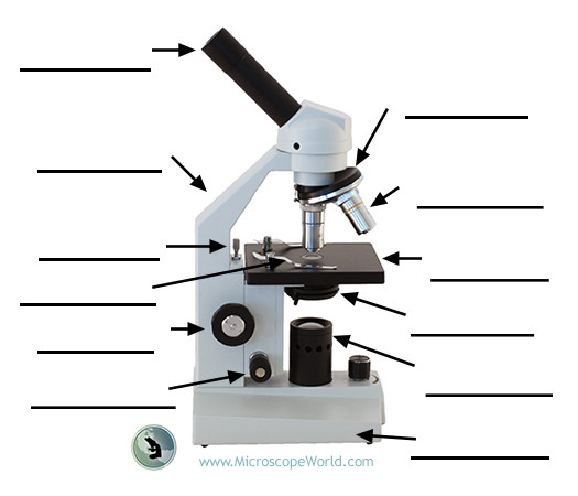
Labeling the Parts of the Microscope | Microscope World Resources Labeling the Parts of the Microscope This activity has been designed for use in homes and schools. Each microscope layout (both blank and the version with answers) are available as PDF downloads. You can view a more in-depth review of each part of the microscope here. Download the Label the Parts of the Microscope PDF printable version here.
16 Parts of a Compound Microscope: Diagrams and Video Once you have an understanding of the parts of the microscope it will be much easier to navigate around and begin observing your specimen, which is the fun part! The 16 core parts of a compound microscope are: Head (Body) Arm. Base. Eyepiece. Eyepiece tube.
Microscope, Microscope Parts, Labeled Diagram, and Functions Microscope, Microscope Parts, Labeled Diagram, and Functions What is Microscope? A microscope is a laboratory instrument used to examine objects that are too small to be seen by the naked eye. It is derived from Ancient Greek words and composed of mikrós, "small" and skopeîn,"to look" or "see".
Microscope Labeled Pictures, Images and Stock Photos photosynthesis. Diagram of the process of photosynthesis, showing the light reactions and the Calvin cycle. photosynthesis by absorbing water, light from the sun, and carbon dioxide from the atmosphere and converting it to sugars and oxygen. Light reactions occur in the thylakoid. Calvin Cycle occurs in the stoma. Neutrophil vector illustration.
22 Parts Of a Microscope With Their Function And Labeled Diagram 22 Parts Of a Microscope With Their Function And Labeled Diagram Microscope Description A microscope is a laboratory instrument used to examine objects that are too small to be seen by the naked eye. In other words, it enlarges images of small objects.
Compound Microscope Parts, Functions, and Labeled Diagram Compound Microscope Parts, Functions, and Labeled Diagram Posted by Fred Koenig on Nov 18th 2020 Compound Microscope Parts, Functions, and Labeled Diagram Parts of a Compound Microscope Each part of the compound microscope serves its own unique function, with each being important to the function of the scope as a whole.
Microscope Labeling Game - PurposeGames.com About this Quiz. This is an online quiz called Microscope Labeling Game. There is a printable worksheet available for download here so you can take the quiz with pen and paper. This quiz has tags. Click on the tags below to find other quizzes on the same subject. Science.
Label Microscope Diagram - EnchantedLearning.com arm - this attaches the eyepiece and body tube to the base. base - this supports the microscope. body tube - the tube that supports the eyepiece. coarse focus adjustment - a knob that makes large adjustments to the focus. diaphragm - an adjustable opening under the stage, allowing different amounts of light onto the stage.
Microscope Diagram Labeled, Unlabeled and Blank | Parts of a Microscope ... Mar 28, 2016 - Print a microscope diagram, microscope worksheet, or practice microscope quiz in order to learn all the parts of a microscope. Pinterest. Today. Explore. ... Coloring Ideas Labeled Microscope Drawing Page - Coloring Ideas. Stephanie Vella. Brownie craft ideas. Medical Laboratory Science. Science Activities. Science And Nature.
Cat Skeleton Anatomy with Labeled Diagram - AnatomyLearner 29/05/2021 · Cat skeleton anatomy labeled diagram. Now, I will show you all the bones from the cat skeleton with a diagram. If you find any mistakes in this cat anatomy labeled diagram, please let me know. I hope this cat skeletal system anatomy labeled diagram might help you understand and identify all the cat’s bones.
Microscope Labeling - The Biology Corner I keep these instructions on the board for the week as a reminder: 1) Start with scanning (the shortest objective) and only use the COARSE knob . Once it is focused… 2) Switch to low power (medium) and only use the COARSE knob . You may need to recenter your slide. Once it is focused.. 3) Switch to high power (long objective).
Parts of Stereo Microscope (Dissecting microscope) – labeled diagram ... If you would like to learn optical components of a compound microscope, please visit Compound Microscope Parts – Labeled Diagram and their Functions, and this article. How to use a stereo (dissecting) microscope. Follow these steps to put your stereo microscopes in work: 1. Set your microscope on a tabletop or other flat sturdy surface where ...
Microscope Parts and Functions Most specimens are mounted on slides, flat rectangles of thin glass. The specimen is placed on the glass and a cover slip is placed over the specimen. This allows the slide to be easily inserted or removed from the microscope. It also allows the specimen to be labeled, transported, and stored without damage.
Compound Microscope Labeled Diagram | Quizlet Contains the ocular lens Body tube A hollow cylinder that holds the eyepiece. Arm Part that supports the microscope. Stage Supports the slide or specimen Coarse adjustment Knob sed to focus when using the low power objective lenses Fine Adjustment Knob Used to focus the image on high power to view image in more detail. Revolving nose piece
Cell Nucleus – function, structure, and under a microscope Immunofluorescence utilizes fluorescent-labeled antibodies to detect specific target antigens. Because of its specificity, you can detect molecules of your interest and see their subcellular localization in cells. Below is an example of the immunofluorescence image. [In this figure] Immunofluorescence staining of nuclei.
A Study of the Microscope and its Functions With a Labeled Diagram ... A Study of the Microscope and its Functions With a Labeled Diagram To better understand the structure and function of a microscope, we need to take a look at the labeled microscope diagrams of the compound and electron microscope. These diagrams clearly explain the functioning of the microscopes along with their respective parts.
Cell Nucleus – function, structure, and under a microscope [In this figure] The cell nucleus diagram and its structure. The nucleus consists of the nuclear envelope like double-layer membranes with pores ( nuclear pores), DNA, nucleolus (a site for ribosome synthesis, plural: nucleoli ), nucleoplasm (like cytoplasm of a cell), and the nuclear matrix, a supportive structure like the cytoskeleton supports cells.
Fluorescence Microscopy- Definition, Principle, Parts, Uses 12/04/2022 · Light Microscope- Definition, Principle, Types, Parts, Labeled Diagram, Magnification; Parts of a microscope with functions and labeled diagram; Organic waste recycling (methods, steps, significance, barriers) Limitations of Fluorescence Microscope. Fluorophores lose their ability to fluoresce as they are illuminated in a process called …
Duck Anatomy - External and Internal Features with Labeled ... Jul 24, 2021 · Here, I will show you again the duck anatomy diagram as a whole so that you may summarize your contents so quickly. I tried to show you the basic anatomical features of a duck. If you need more duck labeled diagrams, please let me know. Or, you may join to anatomy learner on social media to get more updates on duck labeled diagrams.












Post a Comment for "41 microscope labeled diagram"