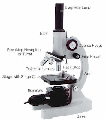
43 labeled diagram of microscope
Simple Squamous Epithelium under a Microscope with a Labeled Diagram ... Web04.03.2022 · Here, in the epidermis of skin labeled diagram, I tried to show you the keratinization over the cell surface. Again, the diagram also shows the cells of the different layers of skin’s epidermis. You will also find the keratinized stratified squamous epithelium on the oral cavity of an animal. Pseudostratified columnar epithelium under a ... Compound Microscope Parts – Labeled Diagram and their … WebMajor structural parts of a compound microscope. There are three major structural parts of a compound microscope. The head includes the upper part of the microscope, which houses the most critical optical components, and the eyepiece tube of the microscope.; The base acts as the foundation of microscopes and houses the illuminator.; The arm …
Compound Microscope- Definition, Labeled Diagram, Principle ... Apr 03, 2022 · Light Microscope- Definition, Principle, Types, Parts, Labeled Diagram, Magnification Amazing 27 Things Under The Microscope With Diagrams 22 Types of Spectroscopy with Definition, Principle, Steps, Uses

Labeled diagram of microscope
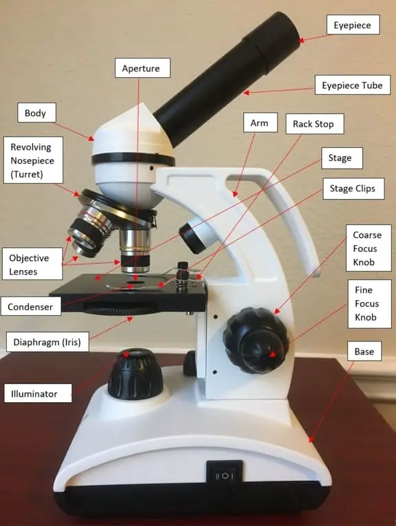
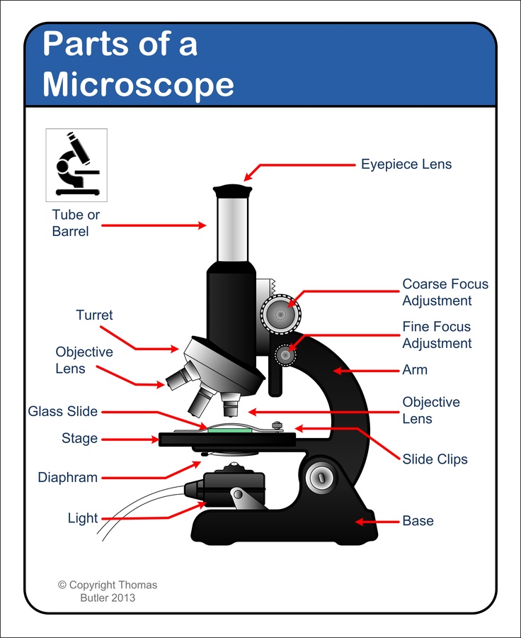
Parts of a microscope with functions and labeled diagram Sep 17, 2022 · Parts of a microscope with functions and labeled diagram September 20, 2022 September 17, 2022 by Faith Mokobi Having been constructed in the 16th Century, Microscopes have revolutionalized science with their ability to magnify small objects such as microbial cells, producing images with definitive structures that are identifiable and ... Inverted Microscope- Definition, Principle, Parts, Labeled Diagram ... Web10.04.2022 · What is an Inverted Microscope? Invented in 1850 by a faculty member of Medical College of Louisiana, named J. Lawrence Smith, this microscope just like it sounds is a light microscope that has its components placed in an inverted order, this means, light source and condenser lens are placed above the specimen stage, pointing down, while … Microscope Parts and Functions WebMicroscope Parts and Functions With Labeled Diagram and Functions How does a Compound Microscope Work?. Before exploring microscope parts and functions, you should probably understand that the compound light microscope is more complicated than just a microscope with more than one lens.. First, the purpose of a microscope is to …
Labeled diagram of microscope. Compound Microscope Parts, Functions, and Labeled Diagram Web18.11.2020 · So, a compound microscope with a 10x eyepiece magnification looking through the 40x objective lens has a total magnification of 400x (10 x 40). Specimen or slide: The object used to hold the specimen in place along with slide covers for viewing. Most slides & slide covers are thin glass rectangles. Stage or Platform: The platform upon … Electron Microscope Principle, Uses, Types and Images ... Oct 03, 2022 · Ans: A light microscope has a low resolving power (0.25µm to 0.3µm) while the electron microscope has a resolution power about 250 times higher than the light microscope at about 0.001µm. Similarly, a light microscope has a magnification of 500X to 1500x while the electron microscope has a much higher magnification of 100,000X to 300,000X. Electron Microscope- Definition, Principle, Types, Uses, Labeled Diagram Web04.04.2022 · Parts of a microscope with functions and labeled diagram; Amazing 27 Things Under The Microscope With Diagrams; Light Microscope- Definition, Principle, Types, Parts, Labeled Diagram, Magnification; Limitations of Electron microscope. The live specimen cannot be observed. As the penetration power of the electron beam is very low, … Cat Skeleton Anatomy with Labeled Diagram - AnatomyLearner Web29.05.2021 · This simple guide might help you to get the basic anatomy of a cat skeleton. I always suggest you read the osteological features of any bones from a cat with a helpful diagram or model. The cat skeletal anatomy labeled diagrams that provide in this article might help you a lot. But if you need more cat anatomy labeled diagrams, please let me …
Parts of Stereo Microscope (Dissecting microscope) – labeled diagram ... Labeled part diagram of a stereo microscope Major structural parts of a stereo microscope. There are three major structural parts of a stereo microscope. The viewing Head includes the upper part of the microscope, which houses the most critical optical components, including the eyepiece, objective lens, and light source of the microscope. Bright-field microscope (Compound light microscope) - Diagram … Oct 04, 2022 · Working Principle and Diagram. Bright-field microscope works on the simple principle of absorption of light. Firstly, the specimen is put on the stage and the light below is focused on the specimen and the specimen absorbs the light and the contrast image that is dark is viewed against a bright background thus creating an image that is magnified and viewed using the objective and ocular lens. Microscope Parts and Functions WebMicroscope Parts and Functions With Labeled Diagram and Functions How does a Compound Microscope Work?. Before exploring microscope parts and functions, you should probably understand that the compound light microscope is more complicated than just a microscope with more than one lens.. First, the purpose of a microscope is to … Inverted Microscope- Definition, Principle, Parts, Labeled Diagram ... Web10.04.2022 · What is an Inverted Microscope? Invented in 1850 by a faculty member of Medical College of Louisiana, named J. Lawrence Smith, this microscope just like it sounds is a light microscope that has its components placed in an inverted order, this means, light source and condenser lens are placed above the specimen stage, pointing down, while …
Parts of a microscope with functions and labeled diagram Sep 17, 2022 · Parts of a microscope with functions and labeled diagram September 20, 2022 September 17, 2022 by Faith Mokobi Having been constructed in the 16th Century, Microscopes have revolutionalized science with their ability to magnify small objects such as microbial cells, producing images with definitive structures that are identifiable and ...
Microscope Maintenance Tips | Science supplies, Microscope ...

The Microscope

Microscope Parts, Types & Diagram | What is a Microscope ...

Phase-contrast microscopy - Wikipedia

Simple Microscope - Parts, Functions, Diagram and Labelling ...
Microscope Parts & Specifications | Microscope World Resources

Living Environment Course

Light Microscope- Definition, Principle, Types, Parts ...

Labeling the Parts of a Microscope: Activity & Lesson Plan ...
SOLVED: 'what to do in this module? Study the diagram of ...

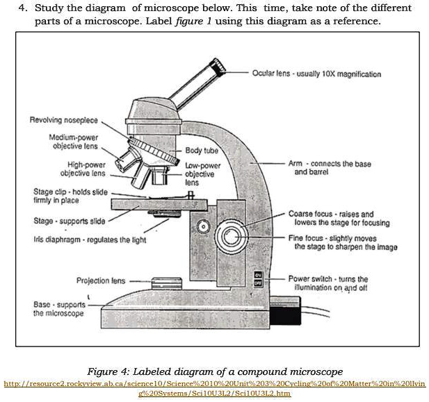
Diagram of a Compound Microscope
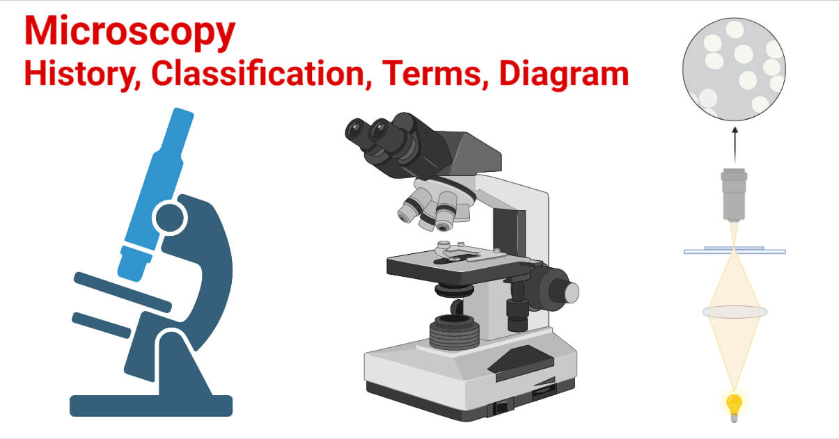
Microscopy- History, Classification, Terms, Diagram
Parts of a Microscope and Their Functions
Diagram of a Microscope - Guide to using a microscope
16 Parts of a Compound Microscope: Diagrams and Video ...
biology labeled microscope diagram - Clip Art Library

give a well labelled diagram of compound microscope using of ...

Parts of a Microscope with Their Functions – Microbe Online

Microscope Labeling Diagram | Quizlet

Microscope Labeling

Label Microscope Diagram - EnchantedLearning.com
Using A Microscope 101: Important Microscope Parts & Functions
Microscope drawing - Teaching resources
Solved Nikon Parts of the compound microscope Write the ...
Microscopes Microscope Parts Quiz on Friday!! - ppt video ...

Parts of the Microscope with Labeling (also Free Printouts ...

Dissecting Stereo Microscope Parts and Functions

The Microscope: Create a Labelled Diagram | Teaching Resources

Compound Microscope Parts, Functions, and Labeled Diagram ...

Compound Microscope Parts, Diagram Definition, Application ...

Microscope | Types, Parts, History, Diagram, & Facts | Britannica

Vektor Stok Microscope Diagram Vector Illustration Labeled ...

Label the microscope — Science Learning Hub
Microscope labelling - Teaching resources

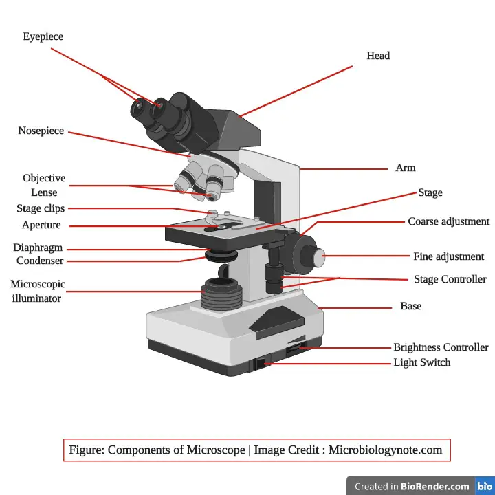
Microscope Parts and Functions

draw a well label diagram of microscope - Brainly.in

Labelling a Microscope Diagram | Quizlet

Diagram of traveling microscope setup with implant cast and ...

Types, Parts and Functions of a Microscope

How to draw compound of Microscope easily - step by step

Parts of a microscope with labeled diagram and functions ...

Biology 4 U no Twitter: "Try this labelled diagram Quiz on ...

5 Important Types of Microscopes used in Biology (With Diagram)
Post a Comment for "43 labeled diagram of microscope"