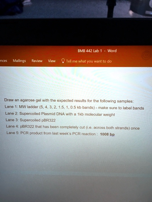
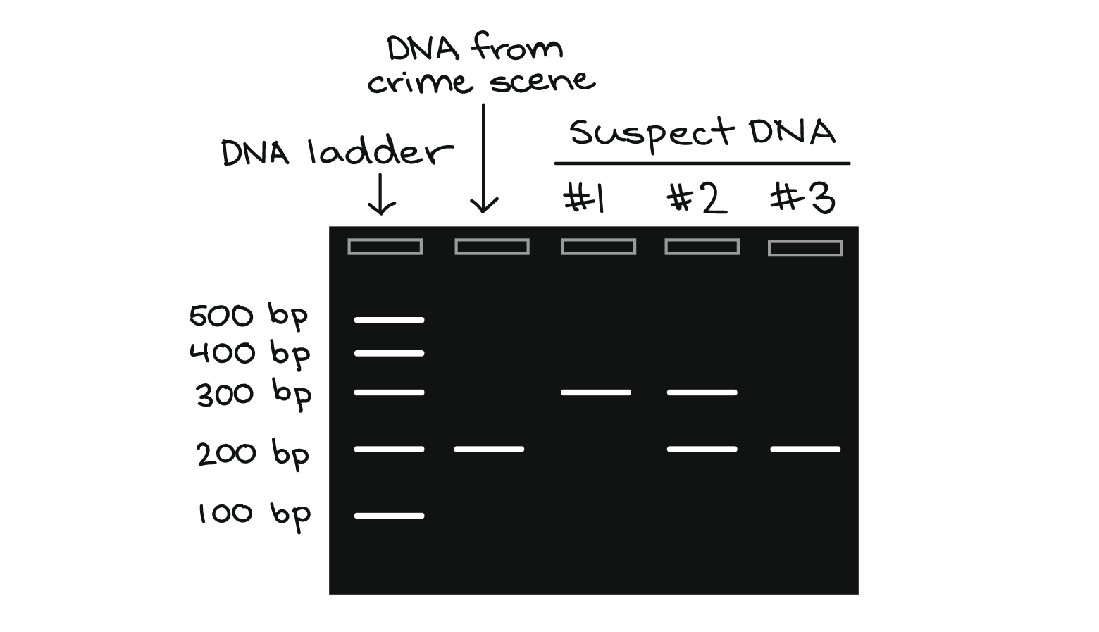
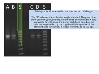
45 how to label gel electrophoresis images on word
PHSchool.com Retirement–Prentice Hall–Savvas Learning Company About a purchase you have made. FAQs: order status, placement and cancellation & returns; Contact Customer Service Analyzing gels and western blots with ImageJ - lukemiller.org Nov 4, 2010 · Obviously this will skew your results if you click in areas that aren’t peaks. If you do happen to click in the wrong place, simple go to Analyze>Gel>Label Peaks to plot the current results, which displays the incorrect values, but more importantly resets the counter for the Results window. Go back to the profile plot and begin clicking ...
Success Essays - Assisting students with assignments online Each paper writer passes a series of grammar and vocabulary tests before joining our team.

How to label gel electrophoresis images on word
Instructions for Authors | JAMA Psychiatry | JAMA Network Photographs, clinical images, photomicrographs, gel electrophoresis, and other types that include labels, arrows, or other markers must be submitted in 2 versions: one version with the markers and one without. Provide an explanation for all labels, arrows, or other markers in the figure legend. US7776521B1 - Coronavirus isolated from humans - Google Patents Disclosed herein is a newly isolated human coronavirus (SARS-CoV), the causative agent of severe acute respiratory syndrome (SARS). Also provided are the nucleic acid sequence of the SARS-CoV genome and the amino acid sequences of the SARS-CoV open reading frames, as well as methods of using these molecules to detect a SARS-CoV and detect infections therewith. Cell Press: Cell Reports Physical Science Table option: Provide the table in the main Word manuscript as described above and embed the scheme within the table where appropriate. In this case, the table (but not the embedded graphic) will be copyedited. Figure option: Combine the scheme and table into a single figure file (e.g., PDF, TIFF, CDX) and label the file as a scheme or figure ...
How to label gel electrophoresis images on word. Primer designing tool - National Center for Biotechnology ... Enter the position ranges if you want the primers to be located on the specific sites. The positions refer to the base numbers on the plus strand of your template (i.e., the "From" position should always be smaller than the "To" position for a given primer). Cell Press: Cell Reports Physical Science Table option: Provide the table in the main Word manuscript as described above and embed the scheme within the table where appropriate. In this case, the table (but not the embedded graphic) will be copyedited. Figure option: Combine the scheme and table into a single figure file (e.g., PDF, TIFF, CDX) and label the file as a scheme or figure ... US7776521B1 - Coronavirus isolated from humans - Google Patents Disclosed herein is a newly isolated human coronavirus (SARS-CoV), the causative agent of severe acute respiratory syndrome (SARS). Also provided are the nucleic acid sequence of the SARS-CoV genome and the amino acid sequences of the SARS-CoV open reading frames, as well as methods of using these molecules to detect a SARS-CoV and detect infections therewith. Instructions for Authors | JAMA Psychiatry | JAMA Network Photographs, clinical images, photomicrographs, gel electrophoresis, and other types that include labels, arrows, or other markers must be submitted in 2 versions: one version with the markers and one without. Provide an explanation for all labels, arrows, or other markers in the figure legend.












-03.1624863098981-21c50c071da0c535da2ef0fcfb6c1e21.png)


Post a Comment for "45 how to label gel electrophoresis images on word"